Wednesday, March 31, 2010

Public lecture and exhibition

6pm, Tuesday, May 4th, 2010, at Culture At Work, 6-8 Scott Street, Pyrmont.
Dr Adam Hamlin will present a lecture on the latest research into Alzheimer's disease. My embroideries will be on display in the gallery. Put the date in your diary now and keep reading the blog or checking the website for more details as the time comes nearer.

Sherryl took this picture of me yesterday, stitching in the studio at 6 Scott Street. It's such a lovely space to sit and sew, even on a drizzly day!

Tuesday, March 30, 2010

Carpet brain

No, carpet brain is not a new nickname for Alzheimer's disease, but the title of the gorgeous image above that I cut out of the Queensland Brain Institute's calendar for 2010 – it's the page for December. Made by Randal Moldrich and Kerstin Pannek, it shows axons in a mouse brain. The caption reads:
Axons, which communicate with other brain regions and the spine, can be modelled using magnetic resonance imaging to indicate direction and intensity.
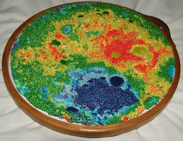
This image immediately appealed to me as inspiration for an embroidery, because I've done something similar before: my map of the far side of the moon in French knots.
This work, which was based on a LIDAR map that NASA made (lidar is radar with lasers rather than radio waves) shows the low areas in blue and high areas in red. In a lovely and improbable twist of fate, the finished embroidery now graces the office wall of a NASA scientist whose daughter bought it from me to give him for his 60th birthday.

As you can see in the picture at the top of this entry, I've outlined a basic shape for the axon map on my black silk fabric. This week I plan to begin filling the space with colourful knots. I remember getting callouses on my fingertips when I made the moon map, so I'll look forward to that!

Monday, March 29, 2010

More cholinergic cells

This rather abstract piece is, once again, inspired by Dr Adam Hamlin's images of apoptosis in the basal forebrain.

Starting with a piece of black silk fabric, I simply began making stitches more or less at random. The thread is Gumnut Yarns Jewels pearl silk thread in Emerald Light. I started with French knots for cell nuclei and long, straight stitches for processes (that's a word I learned in the lab at QBI: it means the structures that look like fibres emerging from the cells).

I didn't plan the design: I am just allowing it to develop in response to the spaces and textures that I create as I make the stitches, so I have no real idea what it will look like when it is finished. This kind of free-form stitching is more difficult in some ways than when you begin with a planned design or pattern, because you never really know where you will place the next stitch or, indeed, when you will be finished. I hope to make this piece quite dense with stitches, but my ideas may change as the work continues.

Sunday, March 28, 2010

Art at work

One of Dr Adam Hamlin's colleagues at QBI made this arrangement of drawing pins on the wall of his or her cubicle. Does it look familiar?

Saturday, March 27, 2010

Are we alone?

The team from the SETI Institute (Search for ExtraTerrestrial Intelligence) do a fantastic science podcast each week called Are We Alone. This week's episode is Thanks for the Memories, including interviews with several neuroscientists and IT professionals.

The podcast is well worth listening to – I subscribe to it through iTunes and often listen to it on my iPod when I'm on the bus or out walking. I always learn something that keeps my synapses firing!

Immunohistochemistry


So how do we find out what’s happening in the brain? To visual the neurodegeneration that occurs in Alzheimer’s disease, we use a process called immunohistochemstry. A technology that allows us to detect the expression of specific proteins in the brain using antibodies that are manufactured to seek out the unique shape of each individual protein.

The first step is to fix the brain tissue with paraformaldehyde. This serves three purposes. First, it freezes the brain in time, so affectively we get a snapshot of the state the brain was in at the time of fixation, secondly, it gives fantastic integrity to the tissue so it doesn’t fall apart during processing, and thirdly, it forms large formaldehyde bonds, cross-linking the proteins so the antibody can recognize the exact shape of each protein. The next step is to section the brain, the sections used for these images is from 40micron brain sections (that’s 1/25th of a millimeter). To achieve this we freeze the brain to -20C then slice it using an extremely sharp blade attached to a machine called a microtome. From a single mouse brain you end up with around 300 sections, which have to be reassembled back into order on glass slides (see
Queensland Brain Institute post by Melody).

The sections next have to be prepared to accept the antibody. All cells, including brain cells have a lipid (fat) cell wall that the antibody needs to penetrate. To do this we soak the brains ethanol, which breaks down the integrity of the cell wall allowing the antibody to move inside the cell. The specificity of the antibody antigen relationship is then further enhanced by soaking the brains in animal plasma and hydrogen peroxide. This prevents the antibodies from binding to non-specific antigens and endogenous peroxidases. The brain tissue is then incubated with an antibody that recognizes your specific protein of interest. This process usually takes around 48 hours. The antibodies are used at incredibly low concentrations, around 1 part antibody to 100,000 parts of buffer (see picture of antibody preparation in Green glow post by Melody).

Binding secondary antibodies that seek out the primary antibodies then further enhances the signal. If the signal is weak then occasionally a tertiary antibody is used to further enhance the signal. These antibodies have a fluorescent tag attached to them.

The sections are then mounted onto a microscope slide. It’s a bit like doing fine art and jigsaw puzzles, as the brain has to be put back in correct order by firstly sorting out the sections into their correct order then gently manipulating a very fragile 40-micron slice of brain tissue onto a piece of glass. Depending on the amount of tissue this process can take 3 or 4 days to complete. The tissue is then coverslipped in a highly alkaline viscous buffer solution so they can finally be viewed on the microscope.

The microscopes used to visualize the fluorescent signal are an amazing piece of technology, costing as mush as a house. Using lasers to excite the fluorophores the highly powerful CCD digital cameras capture the emission wavelength emitted by the fluorescent tag. Any changes in the expression of a protein of interested manipulated by the experimental design can then be analyzed.

Friday, March 26, 2010

Maze embroidery

As in my first embroidery, fly stitches represent neurons making synaptic connections in the maze. The finished work is a meditation on learning and memory.

As a teenager I read Daniel Keyes' novel Flowers for Algernon, and that charming story has been part of the inspiration for this embroidery. The eponymous Algernon is a mouse whose intelligence is raised by some not-clearly-explained means, following which the experiment is tried on Charlie, an adult human. The book is written as Charlie's journal of the process as his intelligence is raised from well below average to far above that of the scientists doing the experiment:
Burt says PSYCHOLOGY means minds and LABORATORY meens a place where they make spearamints. I thot he ment like where they made the chooing gum but now I think its puzzels and games because thats what we did.

The story raises simple moral questions about the value of human life and the progress of science, about which you can come to your own conclusions if you take my recommendation and read the book for yourself.

Thursday, March 25, 2010

All dressed up

Here's a picture that Adam took of me in the lab at QBI... if you can tear your eyes away from the glamorous coverall, note the shower and eyebath in the background, just in case of accidents with toxic chemicals.

Wednesday, March 24, 2010

Of mice and magnets

One of the things I saw during my visit to the Queensland Brain Institute was a mouse having an MRI (Magnetic Resonance Imaging) scan. The selected subject was, Dr Hamlin told me, a rather elderly mouse that was not part of any other experiment. Marianne, the MRI technician, placed him in a container that looked a bit like a large Tupperware salad bowl with a clear lid, and he happily ran around inside the chamber while a mixture of anaesthetic (similar to the pre-meds that you get when you're about to go into surgery, that make you drowsy and relaxed) was pumped in through a tube. Gradually, the little brown patient slowed his running, then lay down to sleep. When he was quite relaxed, Marianne carefully picked him up and laid him in a special contraption that looked like a long piece of plastic pipe that had been cut in half.

To keep his head still and in position during the scan, at one end of the pipe was a small tongue with a hole in it for the mouse's teeth to slot into. A little cowling fitted over his head, continuing to supply the anaesthetic for as long as the scan lasted. Once the sleeping mouse was strapped into position, the whole pipe (with the anaesthetic tubes attached) was placed inside the 16 Tesla MRI imager and Marianne fired up the computer monitors so we could see it working.

An MRI works, Adam and Marianne explained to me – and if I get this wrong, I hope Adam will correct my mistakes – by producing strong magnetic fields so that the hydrogen atoms in the body of the subject all line up and vibrate in a particular direction. Even using the massive magnetic fields this particular machine generates, a detailed image of a one-centimetre-wide mouse brain still takes around four hours to make, during which time the mouse must be kept perfectly still. Even its breathing interrupts the imaging process.

One computer monitor showed the mouse's respiration as a line rising and falling with every movement of its chest. Not just to monitor the mouse's vital signs, this also enable Marianne to time the imaging process so that it only occurred at the same point in the breathing cycle: she would image the brain for a fraction of a second at a time, between each breath in and out.

After several minutes, the first images of the mouse's brain appeared on another monitor. Looking at the magnified images on the screen, and seeing the details that appeared, it was difficult to remember that the brain in question was such a tiny thing residing in such a tiny creature. The purpose of this experiment, Marianne said, was to try to reduce the length of time required to get the level of detail that the neuroscientists needed in the images. By working together, she and Adam hoped to cut half an hour off the time required to image a live mouse's brain.

Adam and I left Marianne to finish the imaging, replacing our watches and credit cards (which had to be left outside so that the magnet didn't fry them) and returning to the QBI lab. Later in the day, when the imaging process was over, our little furry patient would also be returned, to sleep off the effects of his anaesthetic.

Tuesday, March 23, 2010

Green glow

The hallway pictured here is a walkway on the sixth floor of the Queensland Brain Institute, right outside the lab where Dr Hamlin works to create images such as the ones in his post on apoptosis. (Is it just me, or does Apoptosis make you think of a deceased Egyptian pharoah?)

The QBI building is a remarkable work of art in itself, and I like the way the lime green walls and floors glow in the tropical sunlight like fluorophores under the microscope. While I was visiting last week, Adam explained how he attaches fluorescent markers to the cells, a process he has promised to outline in future blog posts. It's complicated but fascinating and I promise it will make your head spin!

Friday, March 19, 2010

Apoptosis


An early and key feature of Alzheimer’s disease is loss of cholinergic basal forebrain neurons. These neurons play a critical role in learning and memory. What triggers these cells to die remains unknown and forms the basis of our research. We do know that they are dying via a process known as apoptosis or programmed cell death. Apoptosis is a mode of cell death in which the cell plays an active role in its own demise. Apoptosis occurs in all multicellular organisms and happens when a cell gets to the end of its life. Apoptosis differs from necrotic cell death as the disposal of cellular debris does not damage the organism and is recycled to be used in future biochemical processes.
In the nervous system this process is crucial to the correct developmental maturation of the brain. You are born with an incredible number of neurons that are undergoing a battle of survival of fittest. Only cells that wire up correctly and integrate into functional circuitry survive. If you don’t use it you lose it. Cells that fail to successfully integrate into the neural circuitry don’t receive critical growth factors and undergo cell suicide or apoptosis. This developmental pruning is a crucial process in the fine-tuning of the brain. A second great wave a neural rewiring and apoptosis occurs during adolescence when the cortex fully integrates itself with lower forebrain emotional centers. Neural apoptosis occurs not only in development, but also in pathophysiological states such as stroke, glutamate toxicity, and β-amyloid toxicity.
The focus of our research is understanding the mechanism by which β-amyloid (the pathological hall mark of Alzheimer’s disease) triggers apoptosis in cholinergic basal forebrain neurons. The series of images inspiring Melody’s embroideries are taken from images of cholinergic basal forebrain neurons undergoing apoptosis. The process used to visualize these neurons is immunohistochemistry. Look out for my next blog where I will attempt to explain the process of attaching fluorescent antibodies to specific proteins to visualize the brain.

Queensland Brain Institute


Yesterday I visited the Queensland Brain Institute at St Lucia, Brisbane, where Dr Adam Hamlin showed me around the laboratory on the campus of the University of Queensland. The picture above shows the three-storey tall mural in the foyer of the building, created by artist Fiona Hall.

Frocked up in a stunning white lab coat, I watched Adam as he prepared slices of mouse brains for staining and mounting on slides. A mouse brain is less than a centimetre across, and it is frozen and cut into very thin slices using a special blade. The slices are 40 microns thick – about 1/25 of a millimetre – and Adam picks them up using a fine paintbrush to move them from one solution to another.Preparing the slices for staining and mounting takes five days, so I only got to see the beginning of the process. At the end of of that time, Adam will have a set of slides that look something like the ones below. The red stain is attached to amyloid plaques that form in Alzheimer's disease, and under the microscope Adam can see the plaques and the swelling of nerves that are affected by them.


Adam's currently supervising a PhD student, Lavinia, who showed me some gorgeous images of neurons being born, growing and migrating in the brains of mice that had had strokes (ischemia). Then MRI technician Marianne let me watch as she sedated a mouse and popped it into a Magnetic Resonance Imaging (MRI) machine -- making sure I didn't have my watch or my credit cards anywhere near the 16-thousand-Tesla magnet.

At the end of a day at the QBI, I felt I had learned so much new information that my synapses were popping! On the plus side, all that stimulation of my neurons will help keep more of them alive for longer. And I've got a million more ideas for my embroideries.

Wednesday, March 17, 2010

Mice and mazes

Today I started work on the series of three embroideries representing the life and death of cholinergic cells in the brain. Thinking about mice and tails led me to think about mazes:

Tuesday, March 16, 2010

Fluorescence

One of the striking things about images of the brain is the use of fluorescent markers to show particular features under the microscope. My first embroidery was worked in a lime green silk thread that approximated the colour of the fluorescence but did not have the same vibrancy.


The embroidery above uses DMC's Fluorescent Effects threads, which certainly have the vibrancy I was seeking, yet the texture leaves a lot to be desired. The threads are 100% rayon, and they are slippery little suckers. They don't grab the fabric or the neighbouring fibres in a satisfactory way, and the stitches slip and slide as you work them.

The stitch I used here is Knotted Loop Stitch, one I hadn't tried before Sharon Boggon revealed it as the stitch of the week on her excellent blog, Pin Tangle. It's a very enjoyable stitch to work, but I would recommend a thread with a softer texture; next time, I'll stick to silk or cotton.

Monday, March 15, 2010

Medley


I found this Moda Vera Medley yarn in Spotlight on the weekend, and I bought some because it matched so perfectly with Dr Adam Hamlin's image of cholinergic cells in a mouse's brain. I plan to incorporate it into one of my embroideries. I think the kids in Culture At Work's Art Think & Play workshops will have some fun with it too!

Saturday, March 13, 2010

Memory and time

My late grandfather, called Pa by all of his grandchildren, was a man with a sharp mind and a vigorous body until quite late in his life. In his eighties, after my grandmother died, he seemed to shrink and shrivel quickly, both physically and mentally: during his final years, his memory lapses seemed sudden and distressing to those who loved him.

I once visited him in hospital, where I found him anxious to get out of bed, because he was waiting for a train that would arrive any minute. A few careful questions revealed that he believed himself at the station in Katoomba, where he had lived as a youth, and he was preparing to catch the train to Sydney. Several days later, when I visited again, he was still waiting for the same train.

My distress at the thought of my grandfather waiting in his hospital room for a train that would never arrive was slightly alleviated by the realisation that he was not worried by the non-arrival of the train. For him, it was simply always about to arrive: three days had passed for me, but he was still on that station platform and the train was still due any minute.

There was a time, though, when the tricks Pa's memory played did distress him. I had brought my son, his namesake, to visit. Max was then about four or five years old, and he looked a great deal like my father (Pa's son) at the same age. When it was time for us to leave I mentioned that we had to get home because Max's dad would be waiting for us. With tears in his eyes, Pa asked, "Who is his father? Am I not his father?"

How could I explain that Pa's own red-headed son had grown up and begotten children who had produced another generation, when the evidence of his eyes and memory told him otherwise?

Pa's dementia showed me how important memory is to our sense of self, and how its loss can affect families and friends. I am sharing this personal story with you because it is one of my reasons for wanting to be involved in this project. Please feel free to share your experiences of Alzheimer's disease or memory loss in the comments below.

Friday, March 12, 2010

Cholinergic neurons

The image at left is of healthy cholinergic neurons in a mouse's brain. It's the first in a sequence of three that Dr Adam Hamlin has shared with me; the other two images show dying, and dead, cholinergic cells. The death of this type of nerve cell is implicated in Alzheimer's disease, and research at the Queensland Brain Institute is looking at triggers for the death of these cells:
Memory loss in people with Alzheimer's disease can be attributed to several factors. These include a build-up of the neuro-toxin Amyloid beta – the major component of amyloid plaques found in patients with Alzheimer's – and corresponding degeneration of a specific population of nerve cells in the basal forebrain.
QBI neuroscientists make Alzheimer's disease advance - UQ News Online - The University of Queensland

This page from my visual journal (left) is simply a rendering of the image above using coloured pencils. The process of shading, sketching and simulating was a useful meditation, although I don't plan for the next embroidery (indeed, the next three embroideries) to be a direct representation of these brain cells, but more of a series of visual comments on the life and death of cholinergic cells and their role in memory.

Thursday, March 11, 2010

Cell birth embroidery


This is a section of the first embroidery piece. It is complete, I think – for now, at least – mostly because I ran out of that particular colour of silk thread and I don't want to introduce a second colour to the design. I used long-legged fly stitches around the central 'tooth', to represent the baby neurons reaching out their arms (or is that wings?) to make connections with loose thoughts floating around in the brain.

To put it in more scientific terms:
QBI's Dr Elizabeth Coulson said a baby's brain generates roughly double the number of nerve cells it needs to function; with those cells that receive both chemical and electrical stimuli surviving, and the remaining cells dying.
More brain research suggests "use it or lose it" - UQ News Online - The University of Queensland

Wednesday, March 10, 2010

Schrödinger's cat and mouse

Thinking about mice yesterday reminded me of the lovely Gwen Harwood poem about quantum physics, Schrödinger's Cat Preaches to the Mice:
Dead or alive? The case defies
all questions. Let the lid be locked.
Truth, from your little beady eyes,
is hidden. I will not be mocked.

Tuesday, March 9, 2010

Tooth and tail


This is my first draft, so to speak, of an embroidery based on Dr Adam Hamlin's image of neurogenesis in the dentate gyrus of a mouse brain. I started stitching in lime green silk thread on a black silk background, creating patterns of stitches that explore some of the texture that's visible in the image. The shape of this area of the brain gives rise to its name: it looks like a particularly sharp and nasty tooth, hence "dentate". But the shape also reminds me of something else... a mouse's tail.

Remember The Mouse's Tale in Alice in Wonderland? As a child I loved the way the words of Lewis Carroll's silly poem trailed down the page in the shape of a tail, making the pun obvious (and making a nightmare for the typesetter in those days, I dare say).

It makes my embroidery a kind of synechdoche (pronounced sin-neck-da-key): in exploring the shape and texture of a part of the mouse's brain I am exploring the whole mouse, and every mouse.

Monday, March 8, 2010

In our brain

I like to listen to Melvyn Bragg's BBC radio podcast In Our Time, which covers a topic in depth each week, with three expert panelists. This week's program, The Infant Brain, is fascinating.

Saturday, March 6, 2010

Nature and art

The Art Gallery of NSW has a new exhibition called Wilderness, and today I attended a forum where five of the contributing artists discussed their works and their art practice. None of the paintings in the exhibition are traditional landscapes; as the curator said, when putting the show together he was less interested in old-fashioned landscape painting than in artists who engage with nature and ideas of the wild. He selected images, he said, that revealed nature through the mind of the artist.

My attention was particularly caught by the paintings of Mary Scott, a Tasmanian artist, whose striking monochromatic images are of stuffed hummingbirds in a case in the Natural History Museum in London. Her birds are painted larger than life, posed in flight or on perches, a strange juxtaposition of life and death. The flattened visual space and menacing shadows of her monochromatic paintings make you feel like you are in the display case with the tiny preserved birds.

The hummingbird at left is sitting on a feeder in my sister's backyard in Colden, New York. Hummingbirds are so tiny, fragile and elusive: the idea of seeing such a lively animal frozen in time and space in a Victorian wunderkammer in a museum is a little disturbing.

Mary Scott said that she felt the same when she visited the museum to photograph the taxidermied birds in their cabinet. She explained, "I am interested in how we understand, consume and classify knowledge in the modern world." Another exhibiting artist, Julie Fragar, said that painting is "a metaphor for humans trying to make sense of the world."

If you are in Sydney or nearby, do try to see this exhibition. Some images in it will astonish you with their profound beauty, while others will challenge your ideas about nature and perhaps help you to make sense of the world.

Thursday, March 4, 2010

Other denizens of the kingdom

Jessica Palmer's bioephemera blog is one that I read regularly, combining science and art with a great sense of humour. Check it out.

Wednesday, March 3, 2010

Unlearning

When I was studying biology in high school, it was common knowledge that you are born with all the brain cells you will ever have. Risk-taking activities such as drinking alcohol and ingesting drugs, we were warned, were bad because they killed brain cells, which would be lost forever. So when Adam showed me his image of newborn brain cells in the dentate gyrus of a mouse (see yesterday's entry), I was blown away not only by its awesome structure but by the fact that something I had thought true for most of my life is wrong.

Neurogenesis in adults, Adam patiently explained, has only been recognised in the past ten years or so. The hippocampus is an area deep within the brain that is involved with learning and memory, and many new cells are born there throughout one's life. Baby cells are born, but only those that receive the right input will survive; one important trigger of survival is links to other neurons, something that occurs during learning. In other words, use it or lose it!

Appropriately, I am creating my first embroidery for this project with the image of neurogenesis in the hippocampus as inspiration. Finding out about neurogenesis has opened a new realm of knowledge for me; thus (I hope) creating neuronal connections for the baby brain cells in my hippocampus and keeping them alive longer.

Tuesday, March 2, 2010

Welcome

Hello to all our readers. I am Melody Lord, a journalist, editor, embroiderer and textile artist. Dr Adam Hamlin is a researcher at the Queensland Brain Institute whose current area of research is into the causes and progress of Alzheimer's Disease (AD).

Adam and I met several years ago during a series of astronomy lectures at the University of Sydney. I was working on an embroidery based on a NASA image of the far side of the moon. Adam gave me a guided tour of his lab and showed me some of his gorgeous microscope images of cellular structures in rat brains, part of his research, then, on motivation and addiction. Now Culture at Work has given us an opportunity to collaborate on a project that combines Adam's research and marvellous images with my embroidery art.

Above: Cell birth in the hippocampus.

Jim Endersby, in his book A Guinea Pig's History of Biology (Harvard University Press, 2007) writes:
Science is the kingdom of the blind: there are no sighted – or even one-eyed – people, because we have no way of looking directly at reality to assess what it is like.
Science is a series of questions about the state of things; it is mutable and changing. Each new discovery debunks an old one and raises questions about a reality that is still beyond our fingertips, as we grope towards the future. Science is the process of asking questions, in the same way as embroidery is a process: you start with a goal in mind, then inspiration, trial and error, and success or failure take your work in new and unexpected directions. As part of our project, both Adam and I will be sharing the process of our work and, we hope, taking you with us on a journey of learning and creativity.

Welcome to the kingdom of the blind.